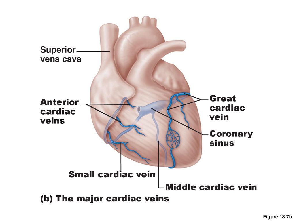
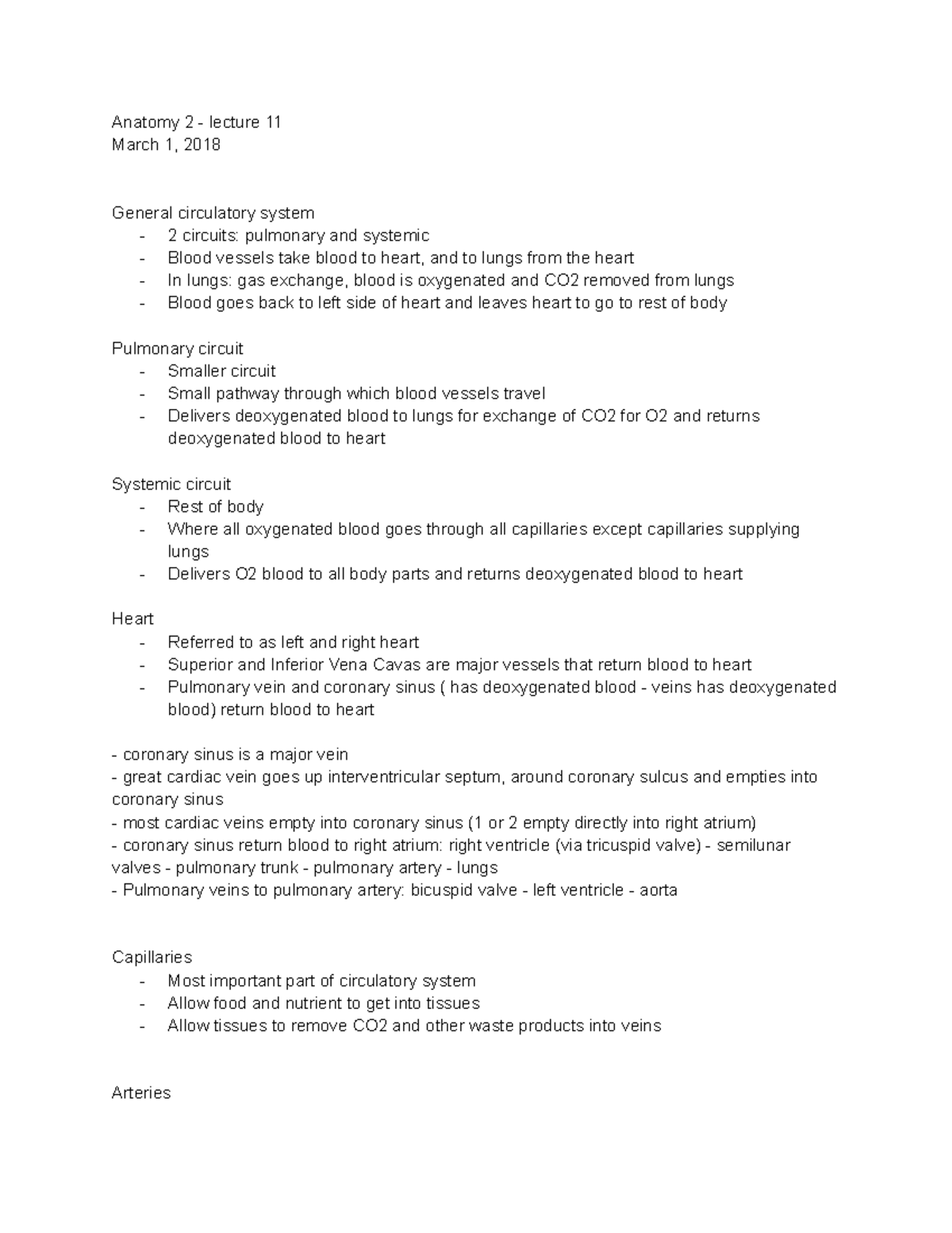
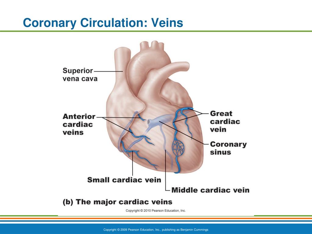
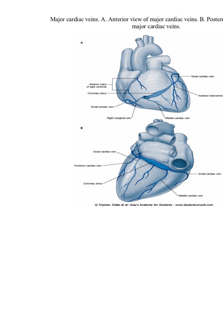
Major Cardiac Veins
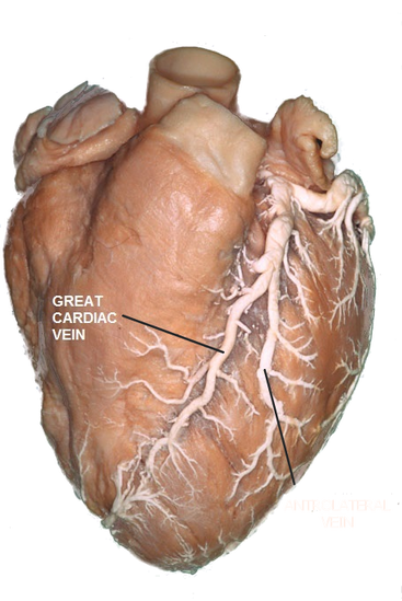
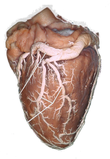
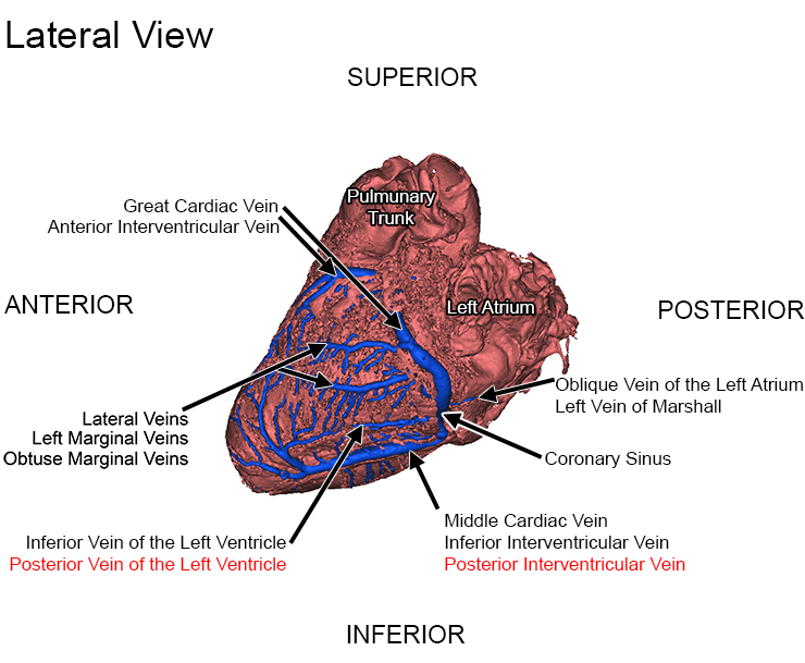
The great cardiac vein originates at the cardiac apex, travels through the anterior interventricular and then to the atrioventricular groove It receives blood from the left marginal vein and other tributaries that drain both ventricles and the left atrium, and empties into the coronary sinus at its origin Middle cardiac vein.
:max_bytes(150000):strip_icc()/heart_and_major_vessels-5820b6ba3df78cc2e887becd.jpg)
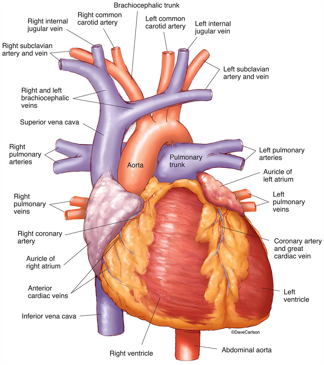
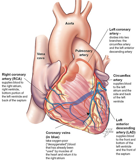
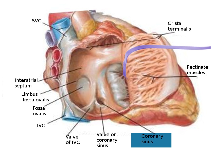
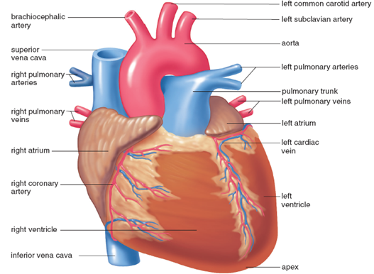
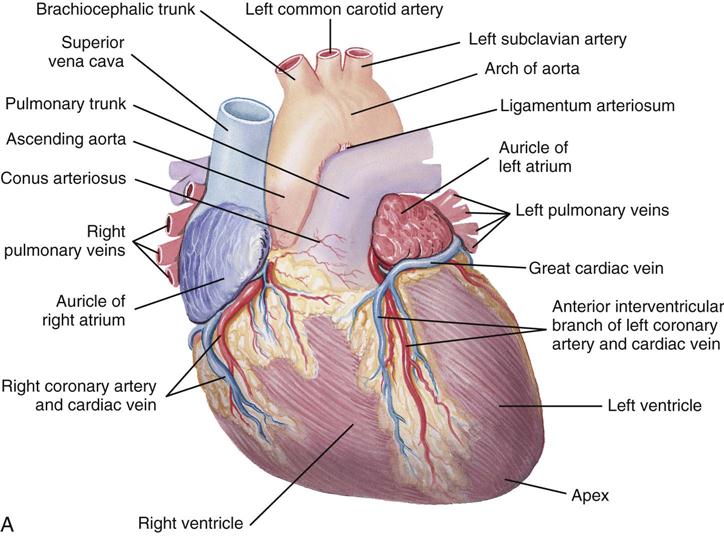
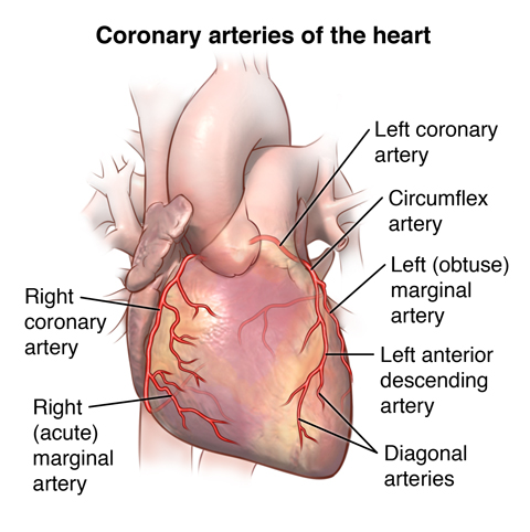
Major cardiac veins. Veins return blood to the heart from all the organs of the body The large veins parallel the large arteries and often share the same name, but the pathways of the venous system are more difficult to trace than those of the arteries Many unnamed small veins form irregular networks and connect with the large veins. The major caudal and right cardiac veins merged into a single trunk in 2 rats (56%) In the remaining case (28%), a common root comprising the major caudal, minor caudal, right cardiac, and cranial cardiac veins was formed before it opened into the caudal part of the right atrium (Figure 4). Heart Blockage – Normal Coronary Arteries There are three arteries that run over the surface of the heart and supply it with blood (see the diagram above) There is one artery on the right side, and two arteries on the left side The one on the right is known as the right coronary.
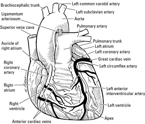
The CS originates at its ostium within the right atrium and extends distally to the valve of Vieussens, where it receives the great cardiac vein Other major tributaries include the left obtuse marginal vein, the posterior left ventricular vein (PCV), the middle cardiac vein (MCV), and the right coronary vein, also known as the small cardiac. Start studying Major cardiac veins Learn vocabulary, terms, and more with flashcards, games, and other study tools. Left coronary artery Originates from left posterior aortic sinus;.
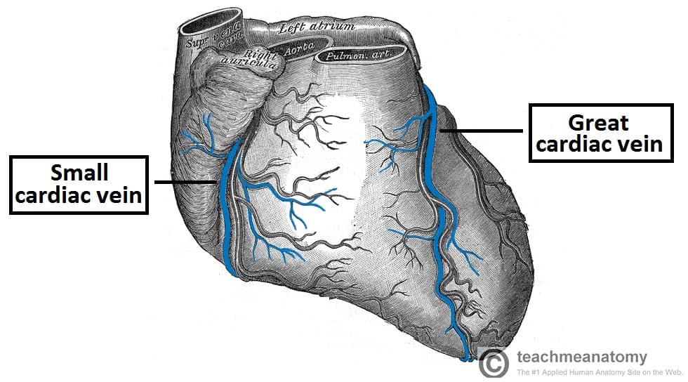
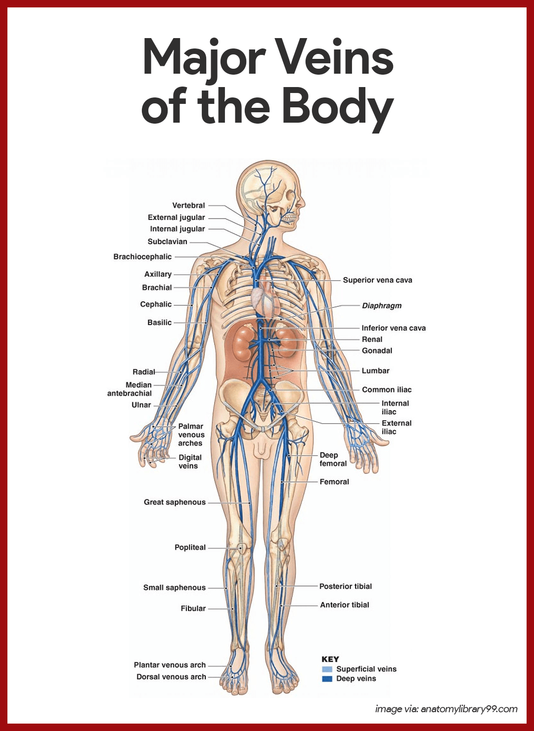
Veins are a type of blood vessel that return deoxygenated blood from your organs back to your heart These are different from your arteries, which deliver oxygenated blood from your heart to the. Major branches sinoatrial nodal, posterior descending, AV nodal, marginal;. The great cardiac vein is the main tributary It originates at the apex of the heart and follows the anterior interventricular groove into the coronary sulcus and around the left side of the heart to join the coronary sinus The small cardiac vein is also located on the anterior surface of the heart.
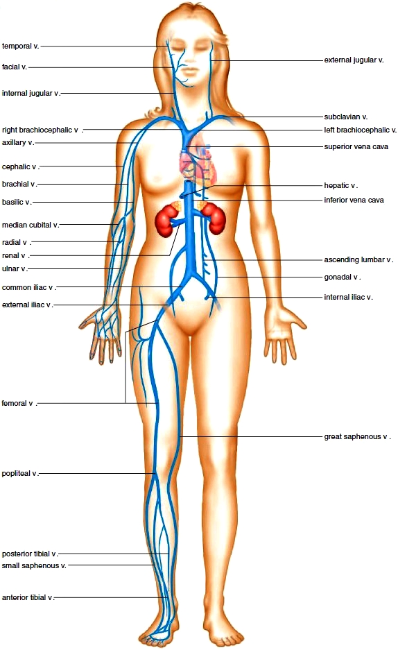
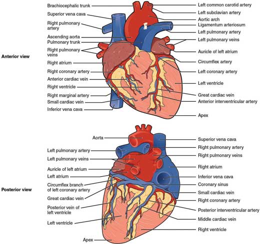
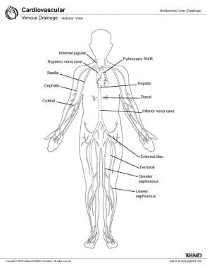
We were talking about the heart again in this week's teaching, so here's an overview of the coronary arteries and cardiac veins. One or more left marginal veins typically merge with the great cardiac vein as it traverses the lateral ventricular wall Small anterior cardiac veinsalso drain blood from the anterior right ventricle directly into the right atrium The anterior cardiac veins 1. Veins of the trunk converge from the thorax, abdomen, and pelvis towards the heart Deoxygenated blood from the thorax ultimately drains into the superior vena cava (SVC) The major thoracic tributaries of the SVC include the azygos venous system, pulmonary veins, internal thoracic vein and cardiac veins.
The left and right coronary arteries branch off from the aorta and provide blood to the left and right sides of the heart The coronary sinus is a vein on the posterior side of the heart that returns deoxygenated blood from the myocardium to the vena cava Hepatic Portal Circulation. Passing anterior to the right coronary artery (RCA), the anterior cardiac veins (ACV) commonly enter into the RA as separate vessels or in some cases (5%–27%) form a common trunk The right marginal vein (RMV) is seen in 80% of cases This vein drains into the small cardiac vein (SCV) in 30% of cases and directly into the RA in 70% of cases. Coronary veins Great, middle, small and oblique cardiac veins drain into the coronary sinus then into the right atrium.
Drain directly into 4 chambers of the heart. Anatomy of the major arteries and veins 3 Anatomy of the heart, the pericardium and valves – FROM CICM 3 Coronary artery anatomy 3 Anatomy of excitatory and conductive elements MAKEUP 4 Electrical properties of the heart 5 Ionic basis of automaticity the normal and abnormal processes of cardiac excitation 5 Pacemaker action potential 5. Representative Results Table 2presents the median anatomical parameters for the major cardiac veins for 42 human heart specimens All heart specimens contained one posterior interventricular vein (PIV) and anterior interventricular vein (AIV).
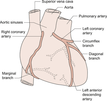
We assessed the prevalence of the major coronary veins for 121 specimens The small cardiac vein was visualized only in 14 of 121 (12%) specimens, which is most likely due to its small size There is also the possibility that the contrast was injected distal to the ostium of the small cardiac vein. Provides blood to the Circumflex artery Runs to the posterior part of the heart, and it provides blood to the left atrium and left ventricle Left marginal artery Runs on the. Major branches left anterior descending, left circumflex;.
Drain into RA 3thebesian veins smallest;. Pathologic cardiac specimens from patients with ccTGA were identified from the Mayo Clinic pathology database Coronary sinus (CS) anatomy and distances from the CS ostium to the major cardiac veins were evaluated Thebesian veins with ostial openings >1 mm, epicardial veins, and venous collaterals were also quantified. The myocardium drains mainly by three groups of veins (1) the CS and its tributaries, which return blood from almost the whole heart;.
Anterior Right Ventricular Venous System Anterior Cardiac Veins The anterior cardiac veins drain twothirds of the right ventricle, including the anterior and Right Marginal Vein The right marginal vein originates near the right ventricular apex and receives branches from the Conus. Arteries vs Veins Arteries and veins are both major components of the cardiovascular system, and both are responsible for transporting blood around the body However, there are several key differences between the two Direction of Blood Flow Arteries carry blood away from the heart, whereas veins carry blood back to the heart. Here are three branches Anterior interventricular artery Runs along the anterior interventricular groove to the apex;.
The great cardiac vein initially parallels the anterior interventricular artery and drains the areas supplied by this vessel It receives several major branches, including the posterior cardiac vein, the middle cardiac vein, and the small cardiac vein. This is a free printable worksheet in PDF format and holds a printable version of the quiz The major cardiac veins By printing out this quiz and taking it with pen and paper creates for a good variation to only playing it online. Answer and Explanation The coronary arteries and cardiac veins are often found together in the grooves of the heart The left coronary artery splits into the left interventricular artery and the.
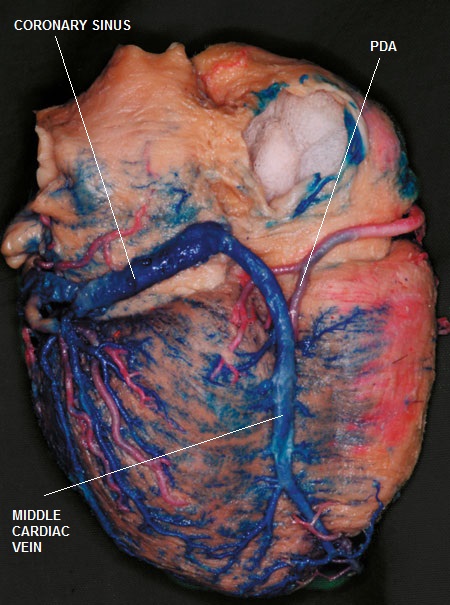
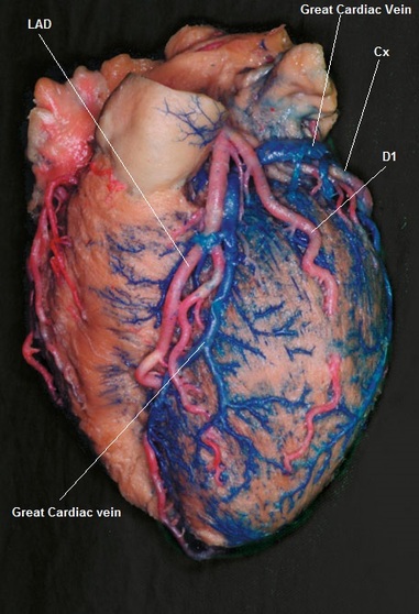
There are two major areas of drainage by the cardiac veins the left side and the right side of the heart The left cardiac vein and the left conal vein belong to the left‐side system, whereas the right cardiac vein and the right conal vein constitute the right‐side system. Coronary Veins Coronary veins drain the heart and generally parallel the large surface arteries (see Figure \(\PageIndex{1}\)) The great cardiac vein can be seen initially on the surface of the heart following the interventricular sulcus, but it eventually flows along the coronary sulcus into the coronary sinus on the posterior surface The great cardiac vein initially parallels the anterior interventricular artery and drains the areas supplied by this vessel. Fig 13 —Major coronary veins draining into coronary sinus Volumerendered CT image from inferior projection shows great cardiac vein receiving left posterior vein and joining in confluence with middle cardiac vein to form coronary sinus, which subsequently drains into right atrium (RA).
Other major tributaries include the left obtuse marginal vein, the posterior left ventricular vein (PCV), the middle cardiac vein (MCV), and the right coronary vein, also known as the small cardiac vein (Fig 253) 16. Large red vessel (the aorta) Large artery that carries blood from of the left ventricle to the arteries of the body Large blue vessel (vena cava) _ (includes the superior and inferior vena cava) _Large vein that empties blood into the right atrium of the heart Cleveland Clinic is a nonprofit academic medical center. The great cardiac vein originates at the cardiac apex, travels through the anterior interventricular and then to the atrioventricular groove It receives blood from the left marginal vein and other tributaries that drain both ventricles and the left atrium, and empties into the coronary sinus at its origin Middle cardiac vein.
Anatomy of cardiac Veins The largest system (Coronary Sinus and it tributaries), which collects a major amount of venous blood from the left The second system (Anterior Right Ventricular veins) which gathers venous blood from the right twothirds of the right The third is the system of the. Representative Results Table 2 presents the median anatomical parameters for the major cardiac veins for 42 human heart specimens All heart specimens contained one posterior interventricular vein (PIV) and anterior interventricular vein (AIV) Some specimens contained more than one posterior vein of the LV (PVLV), posterolateral vein (PLV), left lateral vein (LLV), and/or anterolateral. Major branches left anterior descending, left circumflex;.
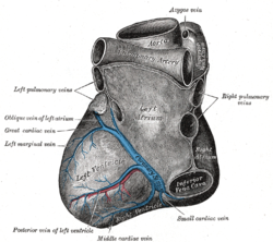
The great cardiac vein, the longest venous vessel of the heart, consists of the anterior interventricular vein and its continuation along the atrioventricular groove 4,7 As the great cardiac vein follows the left atrioventricular groove around the left side of the heart, the great cardiac vein is considered to be in close proximity to the anterolateral commissure of the mitral valve 9. The pulmonary veins carry oxygenated blood from the lungs to the left atrium of the heart Systemic veins carry lowoxygen blood from the body to the right atrium of the heart Capillaries. Your Heart & Blood Vessels The Heart and Blood Vessels Large red vessel (the aorta) Large artery that carries blood from of the left ventricle Front View (Anterior) of the Heart Outside View of the Back (Posterior) of the Heart Coronary veins (in blue) take oxygenpoor ("deoxygenated").
Overview Cardiac catheterization (kathuhturihZAYshun) is a procedure used to diagnose and treat certain cardiovascular conditions During cardiac catheterization, a long thin tube called a catheter is inserted in an artery or vein in your groin, neck or arm and threaded through your blood vessels to your heart. Share your videos with friends, family, and the world. CT cardiac exams provide critical information to practitioners of cardiac therapy Precise information pertaining to the left atrium's complex anatomy, the pulmonary veins, the coronary sinus, or the cardiac veins has a major impact on the efficacy of subsequent cardiac therapy It can speed procedures and facilitate treatment.
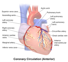
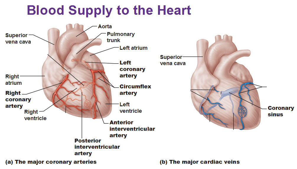
Blood supply to the heart Because of the watertight lining of the heart (the endocardium) and the thickness of the myocardium, the heart cannot depend on the blood contained in its own chambers for oxygen and nourishment It possesses a vascular system of its own, called the coronary arterial systemIn the most common distribution, this comprises two major coronary arteries, the right and the. Some of the most common ones include Deep vein thrombosis (DVT) A blood clot forms in a deep vein, usually in your leg This clot can potentially travel to Superficial thrombophlebitis An inflamed superficial vein, usually in your leg, develops a blood clot While the clot Varicose veins. The major venous vessels of the human heart are coronary sinus, the anterior interventricular veins, left marginal veins, posterior veins of the left ventricle, and the posterior interventricular veins (see also the Coronary System Tutorial).
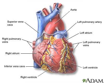
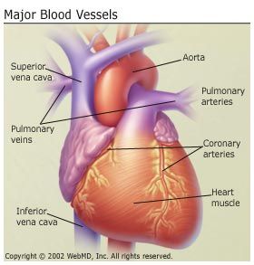
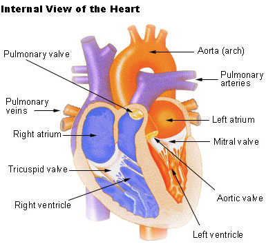
The coronary sinus is formed by three major veins of the heart, the great, middle and small cardiac veins It is approximately 25 cms in length and lies in the atrioventricular groove on the inferior wall of the heart, anterior to the atria, and posterior to the ventricles. Murine cardiac veins consist of several principal branches (with large diameters), the distal parts of which are located in the subepicardium We have described the major branches of the left atrial veins, the vein of the left ventricle, the caudal veins, the vein of the right ventricle and the conal veins. Superior vena cava (from head) Right pulmonary artery Right pulmonary vein Right atrium Inferior vena cava (from body) Right ventricle Aorta Left pulmonary artery Left pulmonary Left atrium Left ventricle Interventricular septum Endocardium Myocardium Pericardium Figure 5–2 Blood flow through the heart.
There were several patterns of major cardiac veins draining the surface of the left artium and both ventricles Cardiac veins return blood to the coronary sinus, directly to the right atrium or to the right cranial caval vein In rodents such as mice the coronary sinus is the terminal. The major cardiac veins in mice do not lie parallel to the branches of the coronary arteries, the latter lying intramurally The results presented above are markedly different not just from those of the human anatomy but also from well‐established descriptions of the heart venous system in larger animals,. And (3) the Thebesian venous network (venae cordis minimae), which is the smaller cardiac venous system and is composed of small venous branches made primarily of endothelial cells that are continuous with the lining.
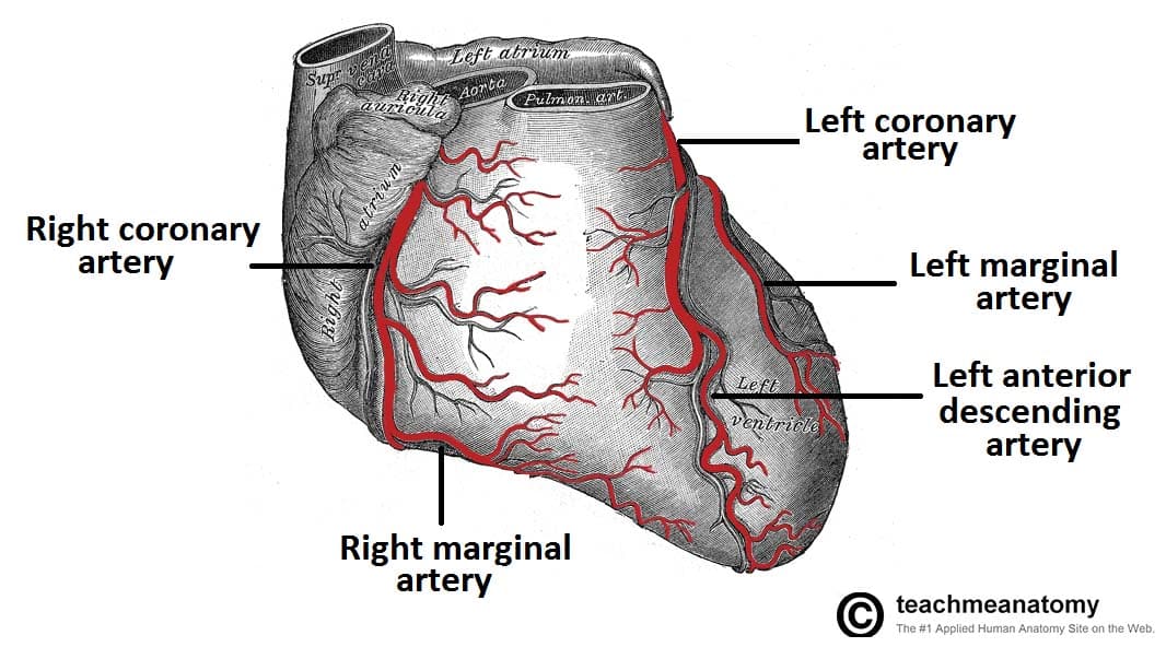
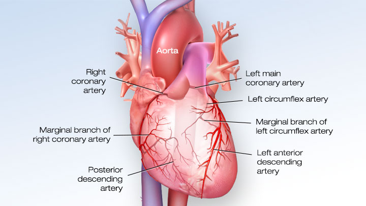
Coronary veins Great, middle, small and oblique cardiac veins drain into the coronary sinus then into the right atrium. Superior vena cava (from head) Right pulmonary artery Right pulmonary vein Right atrium Inferior vena cava (from body) Right ventricle Aorta Left pulmonary artery Left pulmonary Left atrium Left ventricle Interventricular septum Endocardium Myocardium Pericardium Figure 5–2 Blood flow through the heart. The left anterior descending artery branches off the left coronary artery and supplies blood to the front of the left side of the heart The circumflex artery branches off the left coronary artery and encircles the heart muscle This artery supplies blood to the outer side and back of the heart Right coronary artery (RCA).
Start studying Major cardiac veins Learn vocabulary, terms, and more with flashcards, games, and other study tools. Most cardiac veins collect and return blood to the right atrium through the coronary sinus;. We were talking about the heart again in this week's teaching, so here's an overview of the coronary arteries and cardiac veins.
All the veins of the heart (except for those that drain directly into the myocardium) drain into the coronary sinus There are three major veins The great cardiac vein drains the territory of the LAD and circumflex, the middle cardiac vein drains the region of the PDA and other posterior ventricular vessels, and the small cardiac vein drains the anterior wall of the right ventricle and right atrium. ANSWER Two major coronary arteries branch off from the aorta near the point where the aorta and the left ventricle meet Right coronary artery supplies the right atrium and right ventricle with. Vascular disease is any abnormal condition of your blood vessels (arteries and veins) Learn more about the vascular disease types, causes, and treatment.
The 2 main coronary arteries are the left main and right coronary arteries Left main coronary artery (LMCA) The left main coronary artery supplies blood to the left side of the heart muscle (the left ventricle and left atrium) The left main coronary divides into branches. Introduction to the Coronary Veins After flowing through the myocardium, most (80%) of the oxygendepleted blood is returned to the right atrium by several prominent veins that run along the surface of the heart (= epicardial veins) Anterior Veins Draining blood from the anterior ventricles is the great cardiac vein. There may or not be a Thebesian valve covering the ostium of the coronary sinus The major venous vessels of the human heart are coronary sinus, the anterior interventricular veins, left marginal veins, posterior veins of the left ventricle, and the posterior interventricular veins (see also the Coronary System Tutorial ).
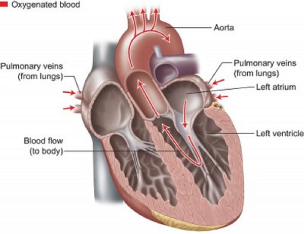
The cardiac veins may be divided into three groups The largest system (Coronary Sinus and it tributaries), which collects a major amount of venous blood from the left ventricle and ends with the coronary sinus, opens into the right atrium. The pulmonary veins carry oxygenated blood from the lungs to the left atrium of the heart Despite carrying oxygenated blood, this great vessel is still considered a vein because it carries blood towards the heart Four pulmonary veins enter the left atrium. Left coronary artery Originates from left posterior aortic sinus;.
(2) the anterior cardiac veins, which primarily drain the anterior regions of the RV and the right cardiac border, ending principally in the RA;. The structure and function of the heart, arteries, veins, and capillaries is vital for the circulatory system to work The overall function of the circulatory system is to transport blood and lymph around the body In doing so it delivers oxygen and nutrients to the body, removes waste products from the body, is involved in the. Anatomy of cardiac Veins The cardiac veins may be divided into three groups The largest system (Coronary Sinus and it tributaries), which collects a major amount of venous blood from the left ventricle and ends with the coronary sinus, opens into the right atrium The second system (Anterior Right Ventricular veins) which gathers venous blood from the right twothirds of the right ventricle and ends in the right atrium.
Major branches sinoatrial nodal, posterior descending, AV nodal, marginal;. BCardiac veins follow same path as arteries include igreat cardiac vein runs with LAD iimiddle cardiac vein follows PDA iiismall cardiac vein runs with RCA ivoblique vein follows post part of LA 2anterior cardiac veins arise on ant surface of RV;.
Edu Cdhb Health Nz Hospitals Services Atoz Publishingimages Pages Education And Development Module 1 anatomy and physiology of the heart 2 Pdf
Cardiovascular System Carlson Stock Art
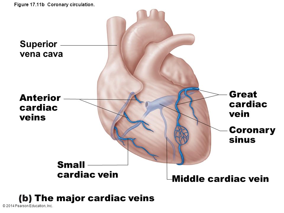
Figure 17 5b Gross Anatomy Of The Heart Ppt Video Online Download
Major Cardiac Veins のギャラリー

Major Blood Vessels Of The Heart

12 December 08 Dote Anatomy Topics

Cardiovascular System Human Veins Arteries Heart

Heart Amboss

Coronary Veins Cardiac Veins
Heart Front View Medlineplus Medical Encyclopedia Image
How The Heart Blood Vessels Work Heart Vascular Institute Temple Health
The Cardiovascular System Ppt Download
Circulatory Routes Major Systemic Arteries Major Systemic Veins
Cardiac Veins Cthsurgery Com
/vascular-system-veins-56c87fa03df78cfb378b3e7c.jpg)
What Is A Vein Definition Types And Illustration
Great Cardiac Vein Wikipedia
Q Tbn And9gcsahyxxrx1jlfi5xp1hb11wm5eriszzvhiimji6bsluaabcb9zv Usqp Cau
Coronary Arteries How It Works Images
Superior And Inferior Venae Cavae
Illustrations Of The Heart Showing The Coronary Venous System And The Download Scientific Diagram

Blood Vessels Biology For Majors Ii
Vasculature Of The Heart Teachmeanatomy

Heart Major Coronary Arteries Cardiac Veins Flashcards Quizlet

Coronary Circulation Of The Heart Bioscience Notes

The Heart And Arteries Veterian Key

Circulatory System
Www Studocu Com En Au Document University Of Melbourne Human Structure And Function Lecture Notes Cvs2 Coronary Circulation And Great Vessels View

Anatomy Of The Heart

Venous Drainage Of The Heart Skudra Net Human Heart Anatomy Drainage Heart Anatomy

Cardiac Veins An Anatomical Review Sciencedirect
Cardiovascular Media Library Watch Learn Live

Coronary Veins Cardiac Veins
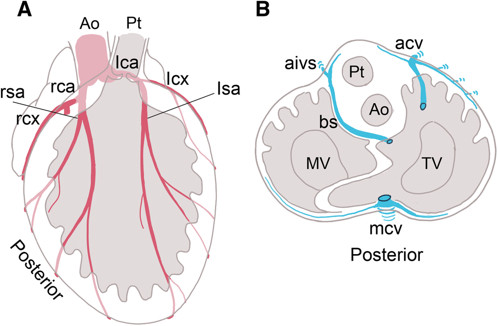
Figure 3 Anatomy Of The Coronary Artery And Cardiac Vein In The Quail Ventricle Patterns Are Distinct From Those In Mouse And Human Hearts Springerlink
Your Heart Blood Vessels
Www Mccc Edu Falkowl Documents Cardiovascularsystem Pdf

Anatomy And Cell Biology 3319 Lecture Notes Fall 17 Lecture 34 Small Cardiac Vein Femoral Triangle Left Coronary Artery

Anatomy Of The Cardiovascular System 3

Quotes About Blood And Veins Quotes
Coronary Arteries And Cardiac Veins Anatomy And Branches Kenhub
Anatomy 2 Study Guide 2 Biol8 Studocu

Coronary Sinus Dr S Venkatesan Md

Heart Anatomy Anatomy And Physiology

Major Cardiac Veins Part 1 Diagram Quizlet
Anatomy Thorax Coronary Sinus Article
Http Www Gmch Gov In Sites Default Files Documents Thorax Heart Blood Supply Innervation Pdf

Pedi Cardiology Anatomy Coronary Veins Coronary Arteries Coronary Arteries Arteries Anatomy Arteries And Veins

Coronary Circulation Anatomy Demonstration Video Medchrometube

The Heart Part 1 Slides By Vince Austin And W Rose Ppt Download
Coronary Arteries Texas Heart Institute
Link Springer Com Content Pdf 10 1007 2f978 3 642 4 6 Pdf

Heart Detail Picture Image On Medicinenet Com

The Coronary Arteries And Major Veins Of The Heart Anterior And Inferior Views Biology Forums Gallery

Coronary Veins Cardiac Veins
Coronary Veins Cardiac Veins
Cardiac Veins Cthsurgery Com

Basic Anatomy Of The Human Heart The Cardio Research Web Project
Vein Wikipedia

Lab 1 Major Cardiac Veins Diagram Quizlet
Nanopdf Com Download Lab Guide Email160protected Pdf
Coronary System Tutorial What Is The Coronary System
Anatomy And Physiology Of The Cardiovascular System Thoracic Key
Body Anatomy Upper Extremity Vessels The Hand Society
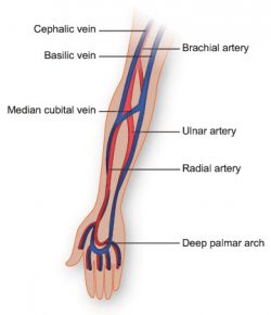
Vasculature Of The Arm Texas Heart Institute
Anatomy And Circulation Of The Heart

Anatomy Of The Heart And Major Coronary Vessels In Anterior Left And Download Scientific Diagram

The Heart

Pedi Cardiology Anatomy Coronary Veins Coronary Arteries Coronary Arteries Arteries Anatomy Heart Anatomy

Cardiac Veins An Anatomical Review Sciencedirect
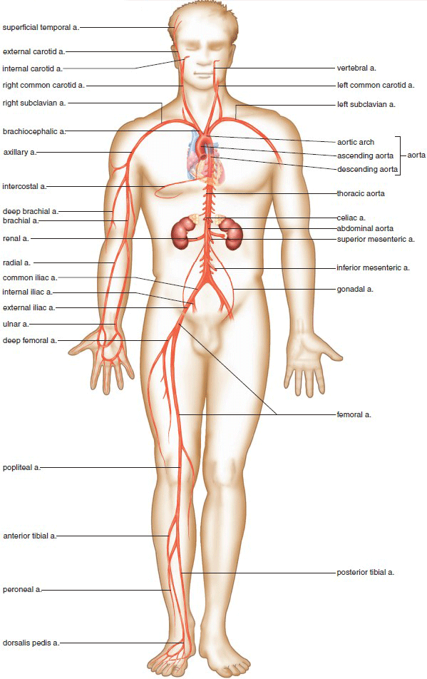
Circulatory Routes Major Systemic Arteries Major Systemic Veins
Q Tbn And9gcs4vuhnoufbevgxb6zoslqk V2sb8kvoeexlpglufaocnbfiuzs Usqp Cau
Ppt Cardiovascular System Powerpoint Presentation Free Download Id 335
Major Differences Difference Between Artery And Vein

Anatomy Of The Heart Coronary Circulation Medical Textbooks Medical Anatomy

Solved 2 Label The Major Arteries And Veins On The Poste Chegg Com
/human-heart-anatomy-1010624306-0115cdf089944788bf5033b8dfb8acf7.jpg)
Aorta Anatomy Function And Significance
Seer Training Structure Of The Heart
Cardiac Surgery Basicmedical Key
Cardiac Veins Cthsurgery Com

Major Blood Vessels Leading To The Heart Superior Vena Cava Inferior Vena Cava Coronary Sinus Video Lesson Transcript Study Com

Arteries Of The Body Picture Anatomy Definition More

Overview Of The Heart Anatomy A Illustration Of The External Anatomy Download Scientific Diagram
Www Heartrhythmjournal Com Article S1547 5271 09 X Pdf

Overview Of The Heart Anatomy A Illustration Of The External Anatomy Download Scientific Diagram

Anatomy Of The Cardiovascular System 3

Coronary Circulation Wikipedia
Major Arteries Veins And Nerves Of The Body Anatomy Kenhub
Lecture 4 Heart Anatomy
Coronary Arteries And Cardiac Veins Anatomy And Branches Kenhub
Venous Drainage Anatomy Overview Microscopic Anatomy Other Considerations
Crossfit The Heart Part 7 Coronary Circulation
Cardiac Veins Cthsurgery Com
Coronary Arteries Wikipedia
Blood Supply To The Heart Thoracic Key
Artery Supply Of The Heart

Arteries Of The Body Picture Anatomy Definition More

16 The Heart Medicine Libretexts
3

Know Ur Heart The Coronary Circulation
Vasculature Of The Heart Teachmeanatomy
Anatomy And Function Of The Coronary Arteries Johns Hopkins Medicine
The Anatomy Of The Human Heart Dummies
Cardiac Veins Cthsurgery Com
Coronary Arteries And Cardiac Veins Anatomy And Branches Kenhub
Q Tbn And9gcs4vuhnoufbevgxb6zoslqk V2sb8kvoeexlpglufaocnbfiuzs Usqp Cau
Cardiovascular System Anatomy And Physiology Study Guide For Nurses


