Image Display In Ct
41 CT Image Display For radiologists, the most important output from a CT scanner is the image itself As was discussed in Chapter 2, although the reconstructed images represent the linear attenuation coefficient map of the scanned object, the actual intensity scale used in CT is the Housfield unit (HU).
Image display in ct. 1 Removes low energy (soft) xrays that do not contribute to image formation but do increase patient dose 2 As the low energy xrays are removed there is a narrower spectrum of xray energies creating a more "monochromatic" beam Image reconstruction is based upon the assumption of a single energy, monochromatic beam 3. The medical images presented on display devices have image pixel values stored as numbers These may be the Hounsfield numbers of a CT image or the numbers sent by a digital radiography device after applying image processed to enhance features. Lookup Tables Following histogram analysis, lookup tables provide a method of altering the image to change the display of the digital image in various ways Because digital IRs have a linear exposure response and a very large dynamic range, raw data images exhibit low contrast and must be altered to improve visibility of anatomic structures.
The pixels of a CT image, for example, are proportional to tissue electron density and are usually expressed in Hounsfield Units, which roughly cover the numerical range 1000 to 1000 A medical image pixel is usually stored as a 16bit integer Two industry standard file formats that support 16bit data are DICOM and TIFF. In some cases CT images are transferred to filmThe camera may be multiformat camera, although most modern CT systems include a laser cameraMultiformat cameras transfer the image display on the monitor to filmLaser cameras bypass the video system entirelyFilm used in CT consists of a single emulsion. 41 CT Image Display For radiologists, the most important output from a CT scanner is the image itself As was discussed in Chapter 2, although the reconstructed images represent the linear attenuation coefficient map of the scanned object, the actual intensity scale used in CT is the Housfield unit (HU).
# MRI scan result on the display of medical computer and projection Similar Images Add to Likebox # MRI scanner room with images from a computerized tomography of Similar Images Add to Likebox # professional brain doctor give a counselor to the patient and. (Computed tomography number) The CT number is a selectable scan factor based on the Houns field scale Each elemental region of the CT image is expressed in terms of Houns field units (HU) corresponding to the xray attenuation (or tissue density) CT numbers are displayed as grayscale pixels on the view ing monitor. Image reconstruction is the term describing the calculation of images from the raw data obtained from the detector modules of the CT scanner This is a process that cannot be performed in real time The reconstruction of image data with a soft tissue or bone algorithm can only be performed from the raw data.
Prices and download plans Sign in Sign up for FREE Prices and download plans. RAD 481 CT Physics and Instrumentation. CT image is a product of complex calculation based on backprojection reconstruction algorithm The CT image is not a real “shade” like in classical Radiography, but a picture which represents with some probability (>95%) the similarity between the real object and its calculated CT image STabakov, 1999.
In CT image display, higher HU values appear brighter and lower HU values appear darker Because the human eye can only distinguish approximately 128 shades of gray, the dynamic range of the CT image display must be adjusted so as to be appropriate to the tissue being evaluated The midvalue of this gray scale is termed the center level, and. Computed tomography is the largest source of medical radiation exposure The benefits of computed tomography are immense and certainly exceed the risks However, this is only true when they are ordered appropriately and studies are optimized to obtain the best image quality with the lowest radiation dose And be sure to take the pledge to Image. CT image is a product of complex calculation based on backprojection reconstruction algorithm The CT image is not a real “shade” like in classical Radiography, but a picture which represents with some probability (>95%) the similarity between the real object and its calculated CT image STabakov, 1999.
This tutorial takes you through the concepts of CT image display including windowing and different types of image reconstructionsThis tutorial has been deve. Image Display Items created automatically by the item recommendation function of My Image Garden, or images saved on the computer are displayed in a slide show (1) Slide Show Area (2) Button Area. At the core of any CT scan image reconstruction is a computer algorithm called Filtered Backprojection (FBP) (Figure 1) Each of the hundreds of xray image data sets obtained by the CT scanner is filtered to prepare them for the backprojection step.
The medical images presented on display devices have image pixel values stored as numbers These may be the Hounsfield numbers of a CT image or the numbers sent by a digital radiography device after applying image processed to enhance features Workstations transform these values to the graphic display values used by the computer graphic card. Computed tomography (CT) is a widely used imaging method However, cardiac CT and CT imageguided therapy are not addressed in this manual table 1 QC test Frequency Gray Level Performance of CT Scanner Acquisition Display Monitors Monthly The radiologist and technologist must look at every study with QA in. CT Image Display STUDY PLAY LCD liquid crystal display most CT monitors today DAC digital to analogy converter CT monitors work with analog signals so data must be converted back to analog to view images window level the 'center' HU of the tissue of interest within the image determines the center of the range.
RAD 481 CT Physics and Instrumentation. 1 Removes low energy (soft) xrays that do not contribute to image formation but do increase patient dose 2 As the low energy xrays are removed there is a narrower spectrum of xray energies creating a more "monochromatic" beam Image reconstruction is based upon the assumption of a single energy, monochromatic beam 3. Display and human inefficiency and variability Statistical tools for measuring performance Next up more interesting phantoms, tasks, etc Thousands of CT images have been made publicly available by CDRH for use in software development & testing •3D tasks and observers •Tasks related to temporal sampling (Fluoro, dynamic CT).
CT Image Display STUDY PLAY LCD liquid crystal display most CT monitors today DAC digital to analogy converter CT monitors work with analog signals so data must be converted back to analog to view images window level the 'center' HU of the tissue of interest within the image determines the center of the range. In this study, the postoperative assessment value of CT postprocessing images was evaluated using multiple imaging techniques, including shaded surface display (SSD), volumerendering (VR), and multiplanar reconstruction (MPR), and was compared with plain radiographs. 1 Acad Radiol 14 Jun;21(6) doi /jacra Optimizing image contrast display improves quantitative stenosis measurement in heavily calcified coronary arterial segments on coronary CT angiography A proofofconcept and comparison to quantitative invasive coronary angiography.
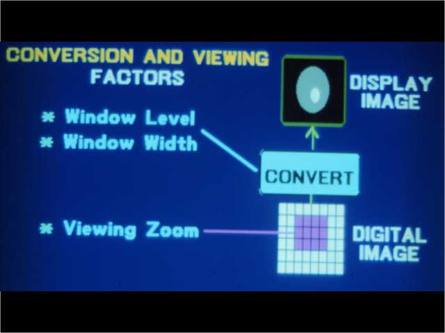
The window width (WW) as the name suggests is the measure of the range of CT numbers that an image contains A wider window width (00 HU), therefore, will display a wider range of CT numbers Consequently, the transition of dark to light structures will occur over a larger transition area to that of a narrow window width (. Interpreting a CT scan Orientation When interpreting at CT scan, it is important to determine the orientation Images are most commonly presented in the transverse plane, and are orientated so that we are looking up the body from the patient’s toes A helpful way to get your bearings is the acronym RALPStarting at the 9 o’clock position and moving clockwise in 90 degree intervals, we. Interpreting a CT scan Orientation When interpreting at CT scan, it is important to determine the orientation Images are most commonly presented in the transverse plane, and are orientated so that we are looking up the body from the patient’s toes A helpful way to get your bearings is the acronym RALPStarting at the 9 o’clock position and moving clockwise in 90 degree intervals, we.
Image reconstruction in CT is a mathematical process that generates tomographic images from Xray projection data acquired at many different angles around the patient Image reconstruction has fundamental impacts on image quality and therefore on radiation dose. Congratulations on making plans to take the Computed Tomography exam As you prepare for the test, this resource will supply you with the information you’ll need to know, including details on the registration process, testing fees, items to bring (and not to bring) on test day, the content you can expect to be assessed on during the exam, and more. Display in socalled stack mode with conventional image display, tile mode, in efficacy of analysis of threedimensional structure on computed tomographic (CT) scans MATERIALS AND METHODS A complex threedimensional phantom composed of entangled tubes was constructed and scanned The resultant images were.
Each elemental region of the CT image (pixel) is expressed in terms of Houns field units (HU) corresponding to the xray attenuation (or tissue density) CT numbers are displayed as grayscale pixels on the view ing monitor White represents pixels with higher CT numbers (bone). As CT technology advances, new challenges arise and solutions emerge to effectively and efficiently produce quality images After completing this program, the participant will be able to 1 Describe the variables that affect contrast resolution in CT images 2 Discuss the various types of artifacts that affect image quality in CT imaging 3. ¾ Display an image in a free viewport ¾ Adjust the window level to the desired value ¾ With the cursor over the image, hold the Shift key and press the Window Level key (F6F11) to save the value to that key.
In scientific visualization, a maximum intensity projection (MIP) is a method for 3D data that projects in the visualization plane the voxels with maximum intensity that fall in the way of parallel rays traced from the viewpoint to the plane of projection This implies that two MIP renderings from opposite viewpoints are symmetrical images if they are rendered using orthographic projection. Threedimensional (3D) medical images of computed tomographic (CT) data sets can be generated with a variety of computer algorithms The three most commonly used techniques are shaded surface display, maximum intensity projection, and, more recently, 3D volume rendering. ¾ Display the image in the primary viewport (blue box) on the Exam Rx or Scan desktop ¾ Select Review Layouts ¾ Select Viewport Format ¾ Select 2 or 4 on 1 Format ¾ With the mouse cursor on one of the images, press F3 for Manual Film Composer or select Shift and F3 for Auto Film Composer How to Change Preset WW/WL keys.
A simple LUT will be a linear "translation" of pixel values to monitor brightness as depicted by the figure below. If you work in the medical field, you’ve likely had to present a patient case report You do a chart review, gather the physical exam and lab data, but often importing the CT scans, ultrasounds, MRIs and other video imaging for display in your PowerPoint can be a timeconsuming and frustrating task I recently discovered. Interpreting a CT scan Orientation When interpreting at CT scan, it is important to determine the orientation Images are most commonly presented in the transverse plane, and are orientated so that we are looking up the body from the patient’s toes A helpful way to get your bearings is the acronym RALPStarting at the 9 o’clock position and moving clockwise in 90 degree intervals, we.
Image Display – Display Data Data are acquired when the xray pass through a patient to strike a detector and are recorded The major components that are involved in this phase of image creation are the gantry and the patient table The gantry and patient table are major components of a CT image. At Image Display, Inc our primary concern is your image ― in fact, your image IS our business Since our start in 1995, we have enhanced the image of hundreds of companies through a successful combination of service, expertise and creative design. 1 Acad Radiol 14 Jun;21(6) doi /jacra Optimizing image contrast display improves quantitative stenosis measurement in heavily calcified coronary arterial segments on coronary CT angiography A proofofconcept and comparison to quantitative invasive coronary angiography.
Computed tomography (CT) is a widely used imaging method However, cardiac CT and CT imageguided therapy are not addressed in this manual table 1 QC test Frequency Gray Level Performance of CT Scanner Acquisition Display Monitors Monthly The radiologist and technologist must look at every study with QA in. If you work in the medical field, you’ve likely had to present a patient case report You do a chart review, gather the physical exam and lab data, but often importing the CT scans, ultrasounds, MRIs and other video imaging for display in your PowerPoint can be a timeconsuming and frustrating task I recently discovered. The medical images presented on display devices have image pixel values stored as numbers These may be the Hounsfield numbers of a CT image or the numbers sent by a digital radiography device after applying image processed to enhance features Workstations transform these values to the graphic display values used by the computer graphic card.
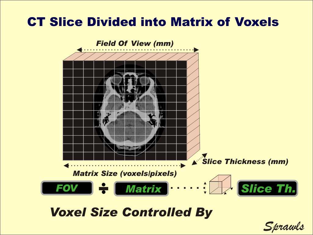
The image matrix is comprised of columns (M) and rows (N) that define the elements or pixels within an image The size of an image is matrix = M x N x k bits The field of view (FOV) is the size of the displayed image. Computed tomography (CT) is a widely used imaging method However, cardiac CT and CT imageguided therapy are not addressed in this manual table 1 QC test Frequency Gray Level Performance of CT Scanner Acquisition Display Monitors Monthly The radiologist and technologist must look at every study with QA in. Display technology, the configuration and maintenance of displays for optimal medical image viewing, and viewing strategies to improve diagnostic performance are discussed The adequate and repeatable performance of the image display system is a key element of information technology platforms in a modern radiology department.
A number of variables should be considered in the evaluation of the accuracy of CT interpretation, including the presence or absence of comparison studies, the specific anatomic region being evaluated, scan complexity, and the mode of image display. "A multimodality display is going to be a color display in order to handle the color in images, such as PET or ultrasound, but it also needs to have the brightness, contrast, resolution, and grayscale calibration to properly display grayscale images such as DR, CR, CT, and MRI". CT image is a product of complex calculation based on backprojection reconstruction algorithm The CT image is not a real “shade” like in classical Radiography, but a picture which represents with some probability (>95%) the similarity between the real object and its calculated CT image STabakov, 1999.
However, 3dimensional rendered CT display of skin and superficial tissues has not seen widespread application Heretofore, physicians have been largely unaware of the diagnostic potential available from this shadedvariant volume rendering of 64slice MDCT. Display of any digital image requires that pixel values (numbers in a computer that range from 0 to some maximum) be allocated a brightness value on a computer monitor This is achieved by use of a look up table (LUT);. Image reconstruction is the term describing the calculation of images from the raw data obtained from the detector modules of the CT scanner This is a process that cannot be performed in real time The reconstruction of image data with a soft tissue or bone algorithm can only be performed from the raw data.
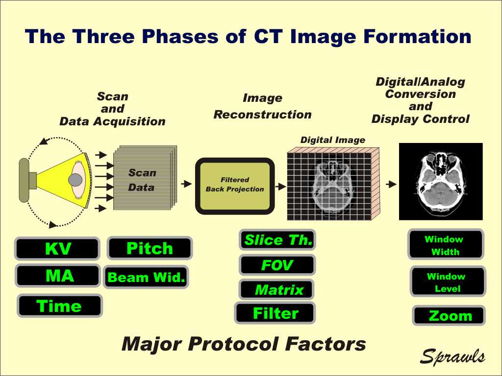
As always, the work done by IMAGE 360 for us was top notch The staff continues to be extremely friendly and helpful I would recommend everyone to use them Steve B, Lyman Hall High School, Wallingford, CT, December. The chamber ionizes the xenon item causing it to migrate to capacitor plate and causing a current in the high voltage load This current is proportional to the radiation and is fed to the computer as data for computing the image The advanced model of CT scanner create image at an angle other than 90° by tilting the gantry Application of CT. The spatial and geometric characteristics of a CT image play a major role in optimizing the imaging protocols That is because the CT image is made up of many small elements or voxels as illustrated here The typical CT image is of a slice through the body During the image reconstruction phase the slice is divided into a matrix of voxels.
Image Display – Display Data Data are acquired when the xray pass through a patient to strike a detector and are recorded The major components that are involved in this phase of image creation are the gantry and the patient table The gantry and patient table are major components of a CT image.

Balpen En Vulpen Parker Jotter Original Ct Display A 45 Stuks Assorti Vullingen Bij Kantoor En Kopie
Connect Slb Com Media Files Core Pvt Lab Product Sheets Coreflow Wholecore Ps Pdf
Ct Compact Technology
Image Display In Ct のギャラリー

Electricity Ct Scan And Mri Scan Display Monitor Adult Screen Size 21 3 Rs Unit Id

Rocktech Display Rk043fn02h Ct Mcu On Eclipse
Video Image Displays For Mr Ct Rt Mr Ct Immersive Patient Experience Creative Healthcare Design By Dti
Q Tbn And9gctrjb70irq5lgefxdbxbhc Gvdjkystoa4zn7yjsnryx7bustfw Usqp Cau

Color Display Unit Ct 5533 Television Panasonic Matsushita
Spie Org Samples Pm1 Pdf
X Ray Image Formation And Contrast

5ctheartscan Ctheartscanphysicians
Ct Series Lowe Rental

Invesalius Display Of The Patient S Ct Scan Results Download Scientific Diagram

Dicom Images Display In Matlab Stack Overflow

Balpen En Vulpen Parker Jotter Original Ct Display A 75 Stuks Assorti Vullingen Ottos

Rise Up Sisters Ct Women S Hall Of Fame

Wholesale Assorted Fidgetz Spinner Display 24 Ct Walmart Com Walmart Com

How To Use The Multi Information Display For The Lexus Ct 0h Lexus Of Reno Youtube
Computed Tomography Ct Daic
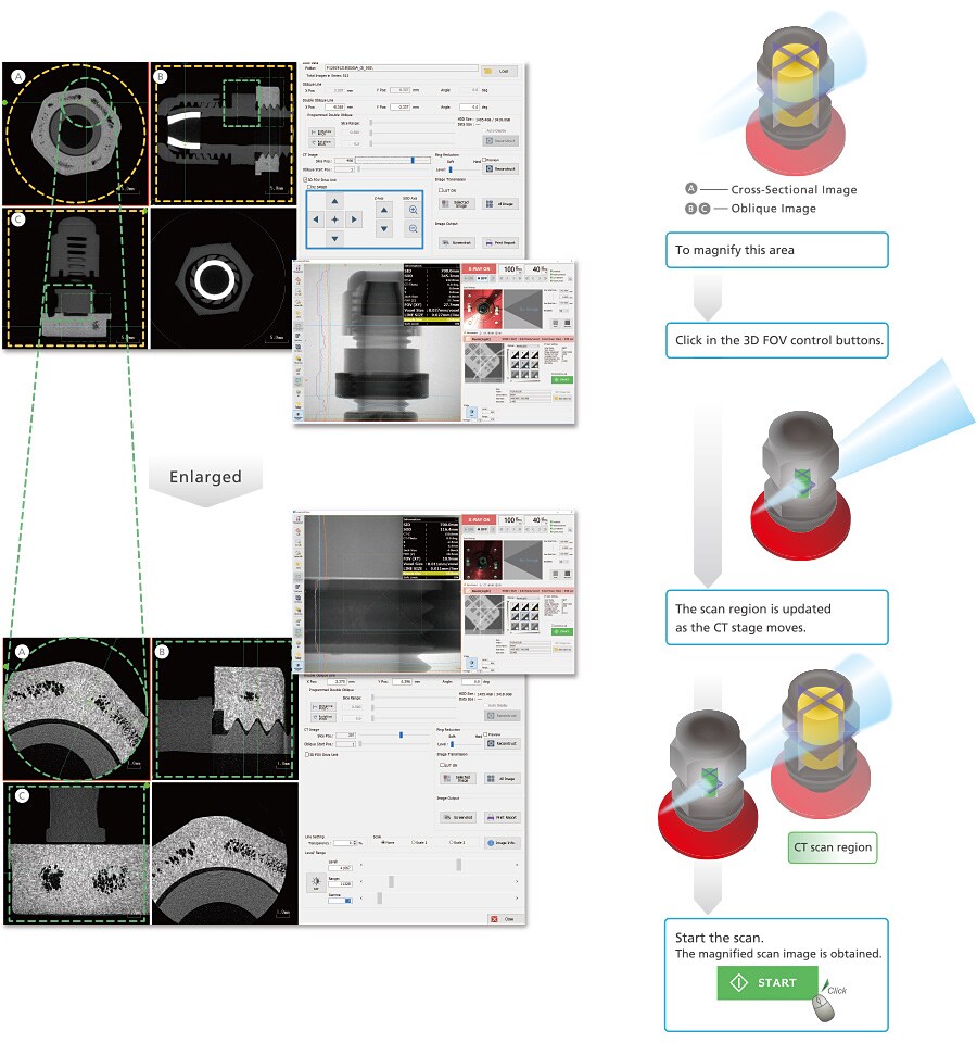
Inspexio Smx 100ct Plus Features Shimadzu Shimadzu Corporation

Histogram Weasis Documentation

Ct Harness Expo Accessories Climbing Technology

Ct Scans And X Rays Display The Damage To The Lungs Of Covid 19 Patients

Wh1604a Yyh Ct Character Lcd Display From Winstar Co 16 Characters X 4 Lines Transflective Positive
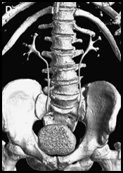
Ssd In Ct Scan Ct Scan Machine

Wh1602b2 Tfh Ct Character Lcd Display From Winstar Co 16 Characters X 2 Lines Transflective Positive Fstn White Background Black Characters

Balpen Parker Jotter Original Ct Display A 45 Stuks Assorti Vullingen Vhk Kantoorartikelen
Flipkart Com Ekta Ct 9914n 14 Digit Display Big Size 2 Power Mode Check Correct Electronic Calculator Ct 9914n 14 Digit Display Big Size 2 Power Mode Check
Ct Body Perfusion Healthcare It Canon Medical Systems

Ct Image Of The Head Displayed On A View Station Of The Pacs The Download Scientific Diagram

Led Diagnosis Display For Ct Mr Imaging China 4m Monochrome Display Made In China Com
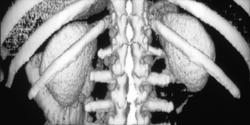
3d Shaded Surface Display Of The Kidney Kidney Case Studies Ctisus Ct Scanning
Ct Image Quality And Dose Management

Balpen Parker Jotter Original Ct Display A 32 Stuks Assorti Vullingen Bij Kantoorexpert

Postprocessing In Maxillofacial Multidetector Computed Tomography Sciencedirect

Ct 22 48v 72v 1v Universal Hall Digital Lcd Programmable Speedometer Display For Electric Motorcycle High Power Bike Hot Deal 09 Cicig

22 Ct Mri 4d Ultrasound Scans In A Holographic Display By Shawn Frayne Through The Looking Glass Medium

Mpr Ct Image Display Demonstrating A Bone Window Mid Sagittal Image Of Download Scientific Diagram

Balpen En Vulpen Parker Jotter Original Ct Display A 75 Stuks Assorti Vullingen Bij Delo
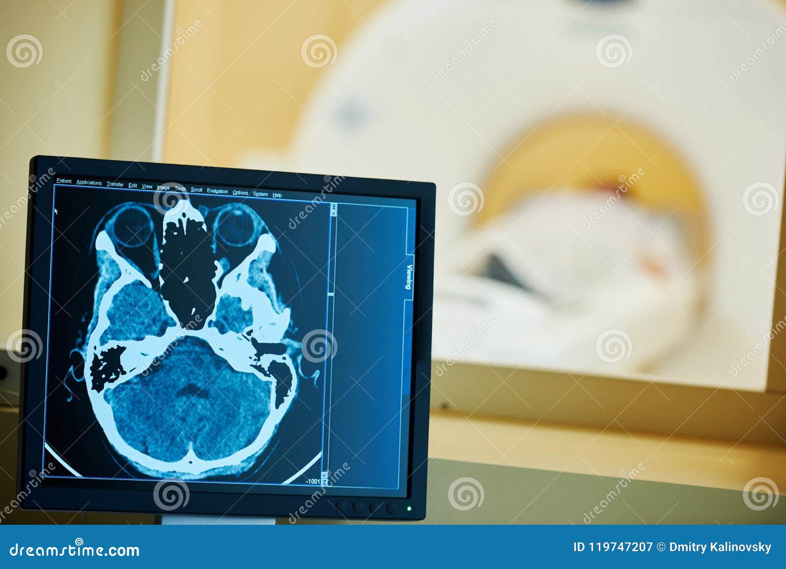
Mri Scan Test Or Computed Tomography Display With Brain X Ray Image Stock Image Image Of Diagnostic Imaging

Pet Ct Viewer Pet Ct Display

Navigatie Display Lexus Ct 0h Hatchback 1 8 16v 2zrfxe 11 Gebruikte Tweedehands Auto Motorfiets En Vrachtwagen Onderdelen Totalparts

Alto Shaam Hsm 24 3s Ct 24 Inch Heated Countertop Display Cabinet
Ct Display Box Idci Parts Network

Balpen En Vulpen Parker Jotter Original Ct Display A 75 Stuks Assorti Vullingen
Http Downloads Lww Com Wolterskluwer Vitalstream Com Sample Content Romans Comprehensive Samples Chap04 Pdf

Ooze Slider Glass Blunt Display 24 Ct Box Msrp 10 99 Each

Ct P5 System Wireless Display Ct D502b Aidbell

Whipple S Winter Wonderland In Killingly Connecticut
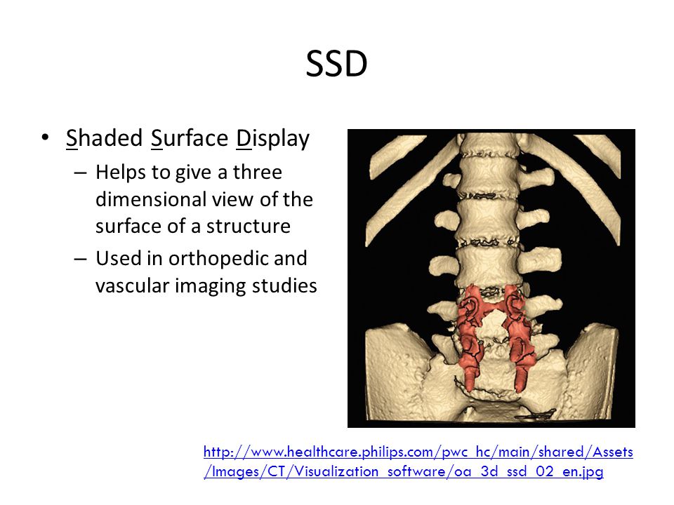
Ssd In Ct Scan Ct Scan Machine

Balpen En Vulpen Parker Jotter Original Ct Display A 45 Stuks Assorti Vullingen
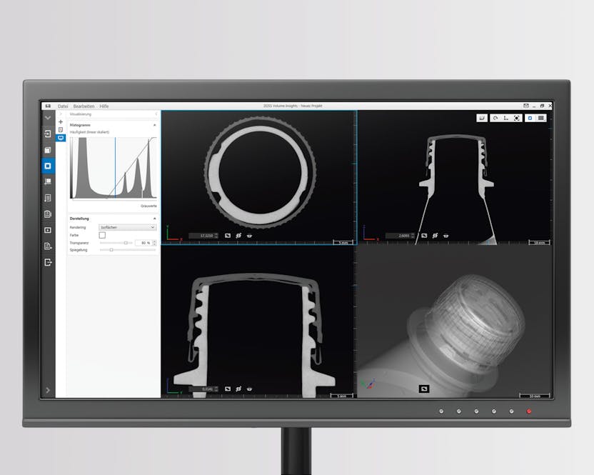
Benefits Of Ct Technology

Vulpen Parker Jotter Original Ct M Assorti Display A Stuks Muldi

Lcd Alphanumeric Display Winstar Wh04a Ygh Ct Gm Electronic Com

Vulpen Parker Jotter Original Ct M Assorti Display A Stuks Kopen Bestel Online Bij Hijdra Com
Ct Image Quality And Dose Management

File Viewer Ct Keosys Jpg Wikipedia
Bv800 Ct Fume Cabinet Digital Display Benchvent
Mr Solutions New Preclinical Imaging Pet Ct Range On Display At Eanm 19 Mr Solutions

Phone Uv Sanitizer Counter Display 12 Ct
Pzem 022 Open En Dicht Ct 100a Ac Digitale Display Power Elektronische Apparatuur Marktplaats Nl
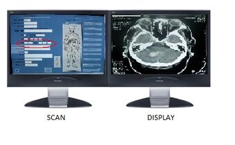
Computed Tomography Htm Wiki Fandom

Buy Tiger Balm Top Sellers Display 14 Ct

Electronics High Quality Large Display Dual Power Ct 512 Wt N Calculator Amazon In Office Products

Ooze Slim Twist Display 48 Ct Categories
Calculator Citizen Ct 933n 12 Digit Dual Power Big Display Shopee Philippines
Trojan Magnum Tub Display 48 Ct Cstore1
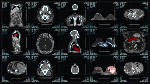
Hospital Mri Examinations Display With Stock Footage Video 100 Royalty Free Shutterstock
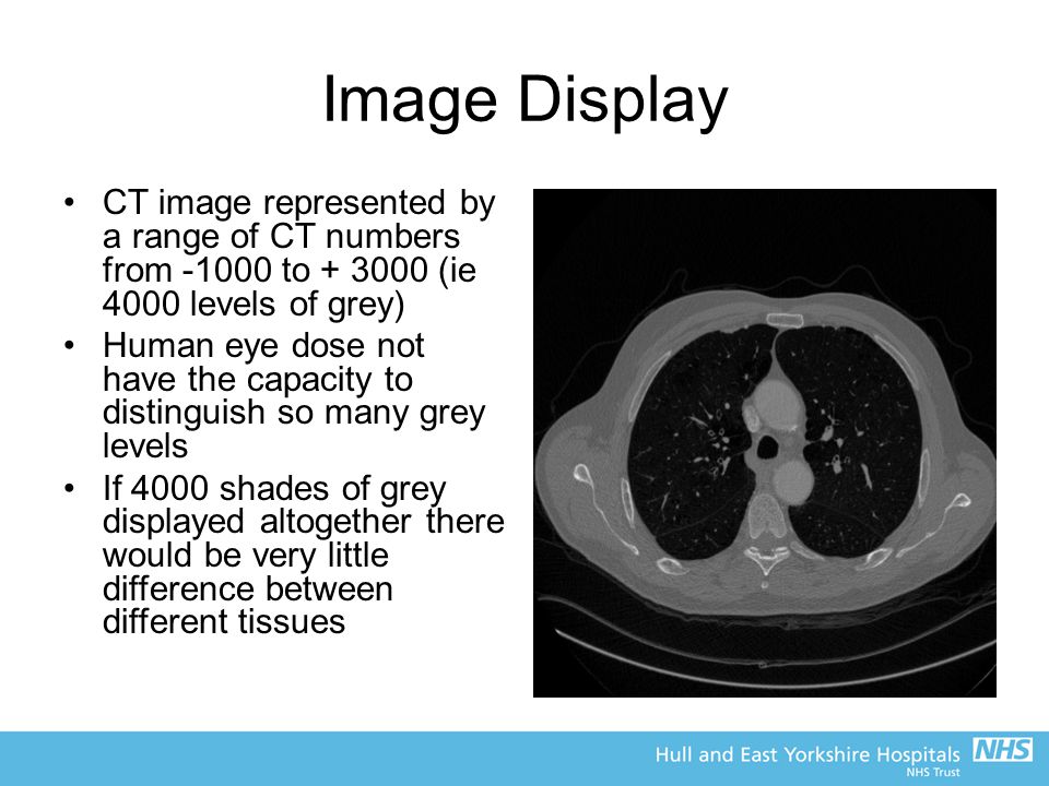
Ct Scanning Dr Craig Moore Ppt Video Online Download
609358.jpg)
Online Balpen Parker Jotter Original Ct Assorti Display A Stuks Kopen Bestellen

Ct Numbers Window Width And Window Level

Contrast Agents For Ct Scans Time To Rethink The Risk Shots Health News Npr

12 Digit Solar Battery Dual Power Large Display Office Desktop Calculator Ct 512 Buy At A Low Prices On Joom E Commerce Platform

Round Digital Device With Ct Display Purple Sphere Circle 02 Ct Purple Sphere Png Pngegg

Display Examples Of The Axial Ct Images On Which Automatically Labeled Download Scientific Diagram
Q Tbn And9gcs7mplwtg551zzkpozjuhf9vkwg0ggs8rsptqfenpekvn5tzhkn Usqp Cau
Q Tbn And9gcsstgjmu7fsc7q Wkr4jfspojomjhzr6jqaxsk Scpfeto1euef Usqp Cau

Ct Image Display And Reconstruction Youtube

Pez Easter Tube Counter Display 18 Ct 594

Bic Zodiac Lighter 50 Ct Display Abcproductsinc

Ct Image Quality

7me6910 1aa10 1ad0 Siemens Signal Converter Mag 5000 Ct I

Single Phase Whole Current Ct Operated Meters With Lcd Display Technitengineering

Huge Ct Halloween Display Depicts Current True Life Horrors

A Connecticut Man S Halloween Display Features Real Life Horrors The Coronavirus Pandemic And Black Lives Lost Cnn

Apm Digital Ct Panel Meter Hoyt Meter
Fujifilm Contemplating Purchase Of Hitachi Medical Systems Imaging Technology News
Ct 912nd Premium Quality Big Display Big Button 12 Digit Big Size Calculator Buy Online At Best Price In India Snapdeal

Diagnostic Monitors For Cross Sectional Ct And Mri Images

Balpen En Vulpen Parker Jotter Original Kopen Mkb Office Shop

Hyde Color Edition 70 Ct Display Disposable Device And Pods Usa

Connecticut Man S Holiday Display Grows To 3 Dozen Inflatables To Share Love For The Holidays And Promote Giving Hartford Courant

Christmas Floor Display 144 Ct My Neighborhood Mattress

Wireless Display Ct D502 Aidbell

Afstandsbediening Voor Toshiba Ykf326 024 Ct 8056 Td Z472 Lcd Display Monitor Afstandsbedieningen Aliexpress

Ct 19 Machinecontrol Duo Set Kilverbesturingssysteem In Koffer Omtools

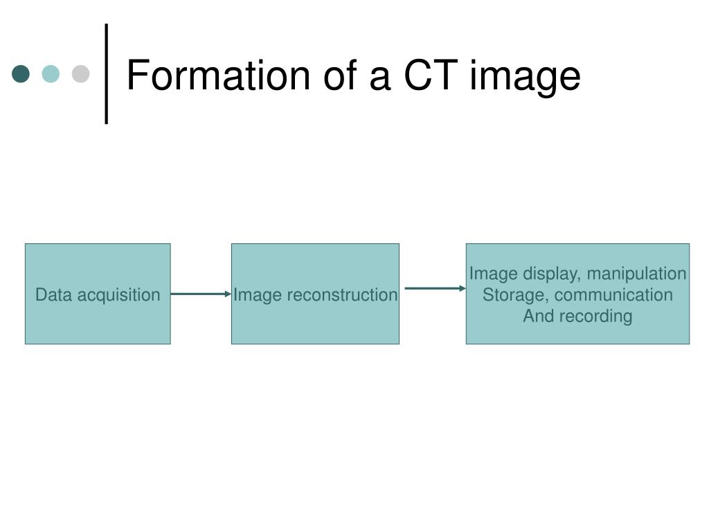
Computed Tomography Formation Of A Ct Image Data Acquisitionimage Reconstruction Image Display Manipulation Storage Communication And Recording Ppt Download

Balpen Parker Jotter Original Ct Display A 32 Stuks Assorti Vullingen Wuestman
Q Tbn And9gctwhfhsryl0ctt2 Qr54osmiun3n9d3kofnz Soorcs44dmvlet Usqp Cau
How We Read Fch Pet Ct For Prostate Cancer Cancer Imaging Full Text
Principles Of Ct
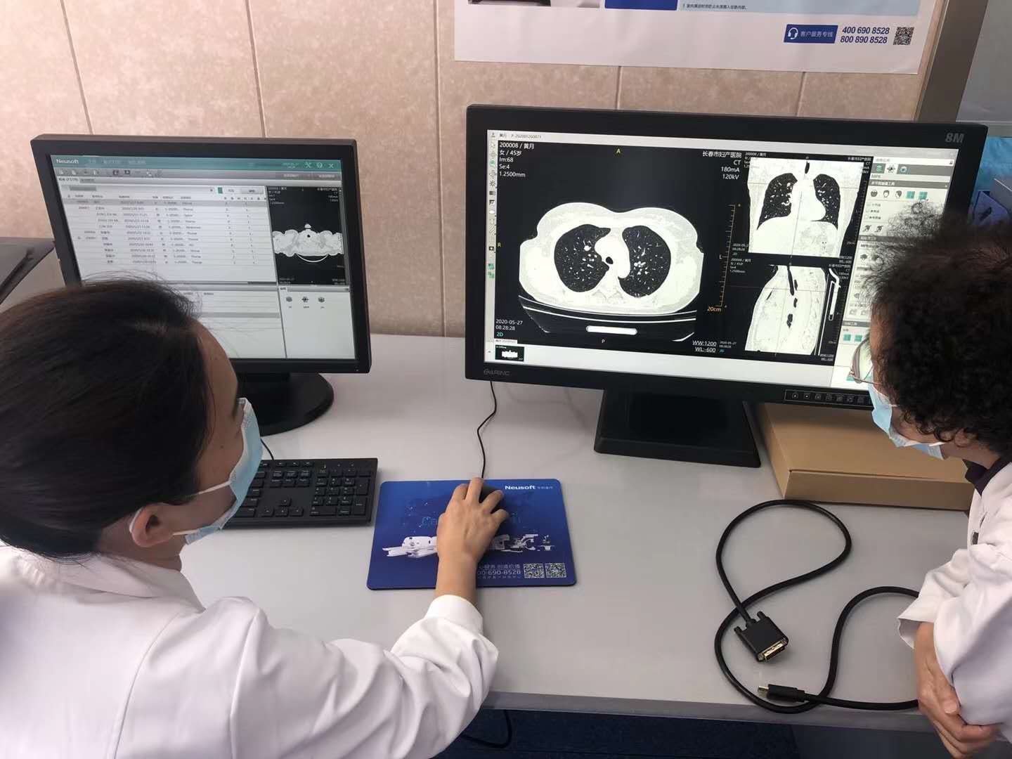
Led Diagnosis Display For Ct Mr Imaging China 4m Monochrome Display Made In China Com

Phone Charger Display 172 Ct Nimbus Imports
Ppt Computed Tomography Powerpoint Presentation Free Download Id


